Complete rehabilitation of an unsatisfactorily restored mandible
Two-piece implants and the alveolar ridge-splitting technique
Case description
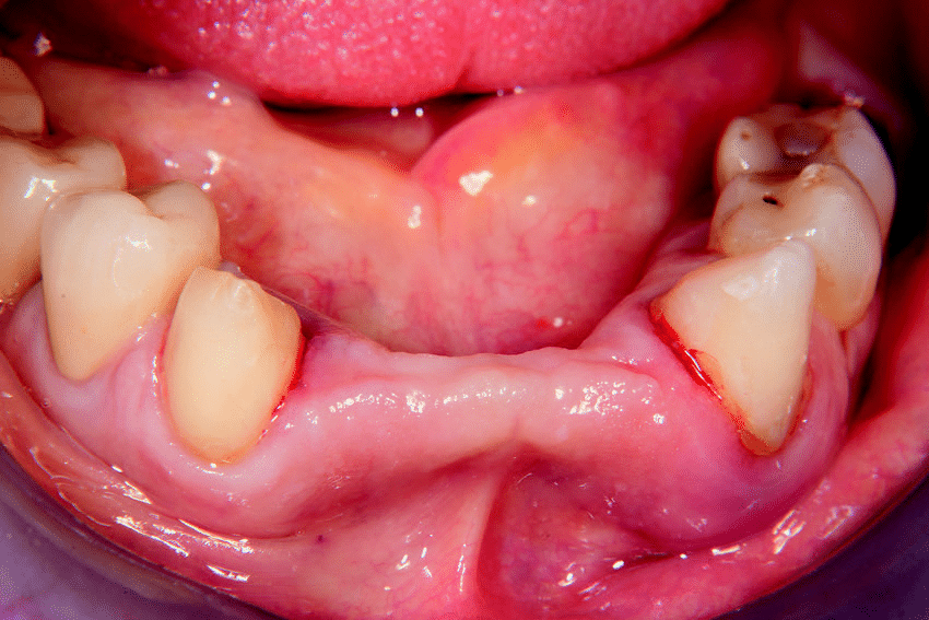
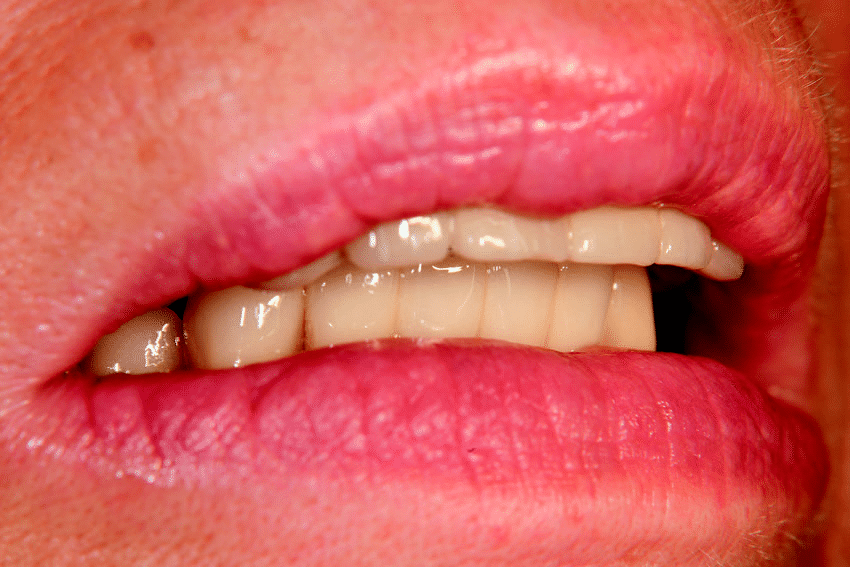
A 65-year-old female patient presented herself with insufficient fillings, crowns and bridges in the mandible (Fig. 1). The first molar on the left side showed advanced mobility (Grade III). The bridgework in the anterior region was to be replaced by single-tooth crowns on the remaining natural teeth and a bridge supported by two implants in regions #32 and 42 was planned. By using the bone-splitting technique, the local bone in this area was to be enhanced (Figs. 2–5). After removal of the hopeless teeth #36 and 37 and subComplete rehabilitation of an unsatisfactorily restored mandible Two-piece implants and the alveolar ridge-splitting technique
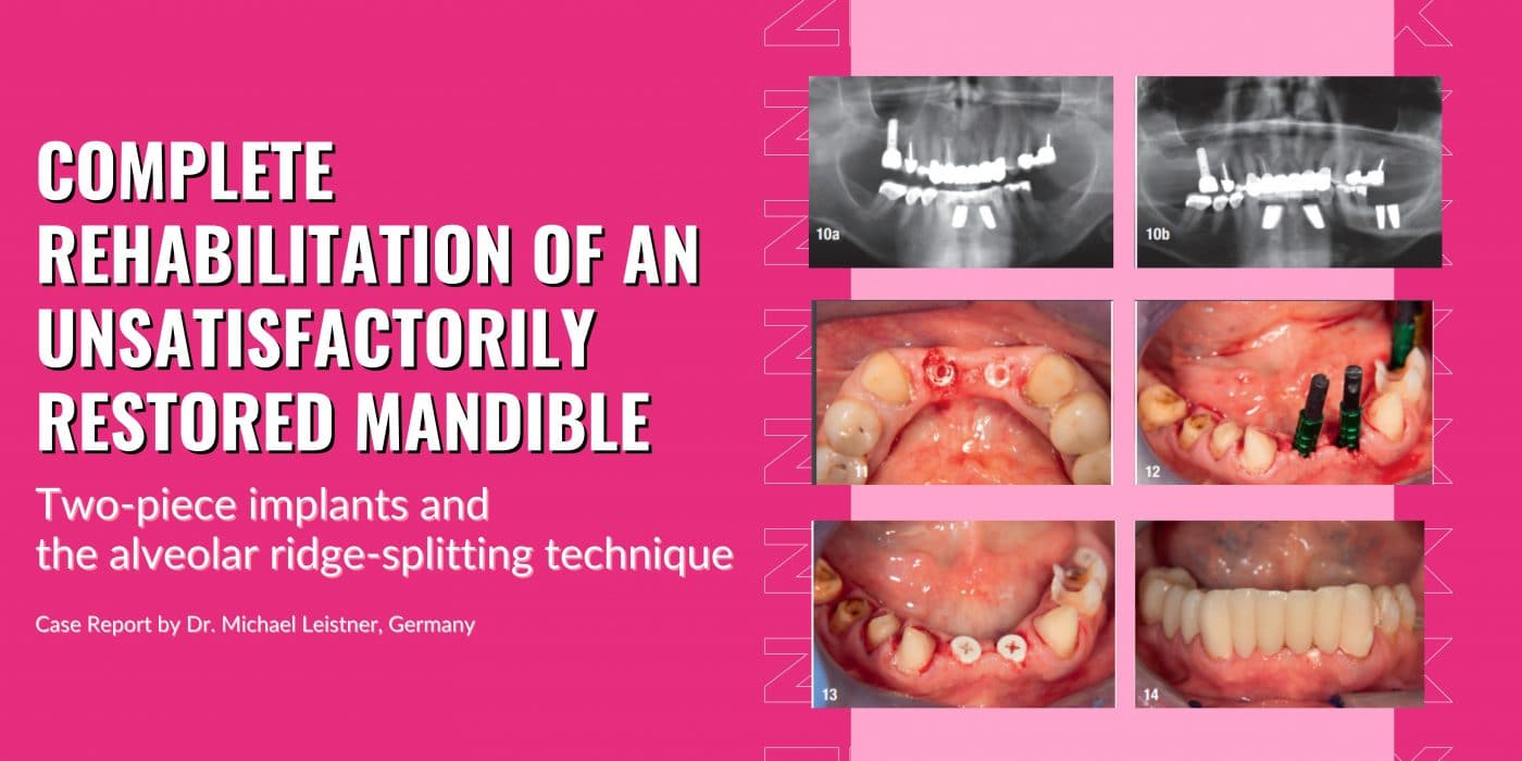
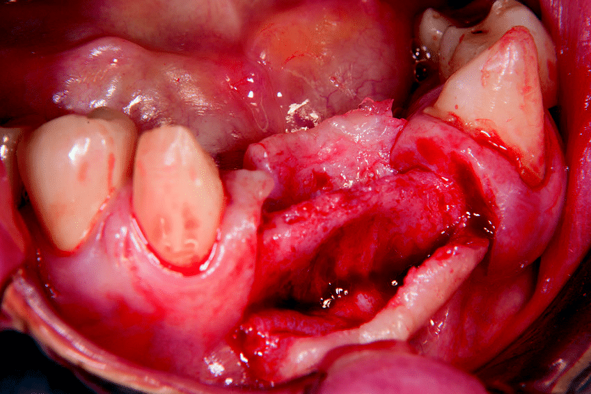
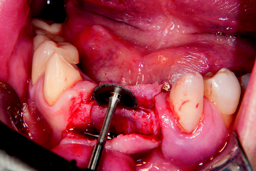
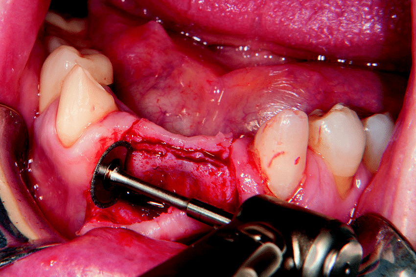
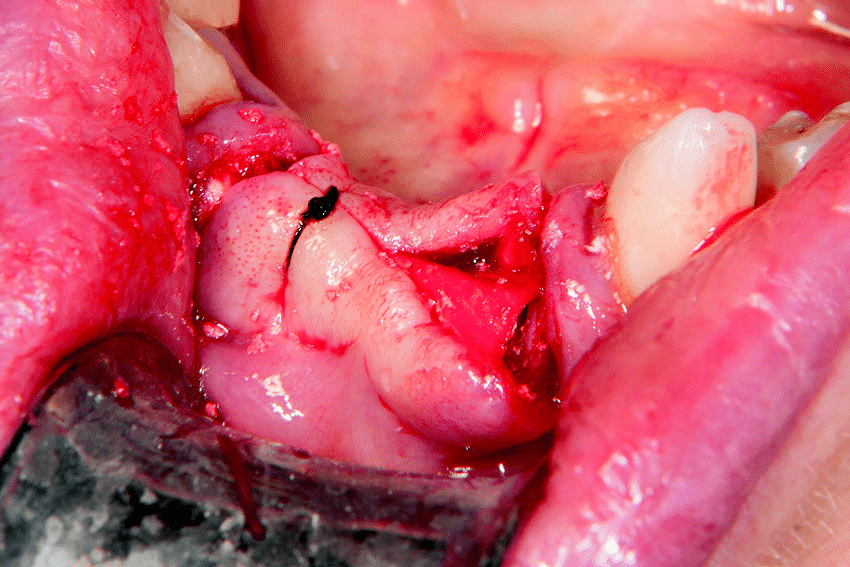
Fig.1: Initial situation: radiograph (a) and clinical view (b). Fig.2: Intra-oral situation after tooth removal and completed tissue healing. Fig.3: Opened and mobilised flap and exposure of the residual bone. Fig.4: Sagittal cut to split the bone ridge using a small fine-grained diamond disc. Fig.5: Vertical cuts at the outer limits of the bone flap. Fig.6: Situation after spreading of the bone, implant placement and filling of the remaining defects with autogenous bone chips. Fig.7: Subsequently, covered with a removable membrane, the flap is repositioned and fixed with sutures.
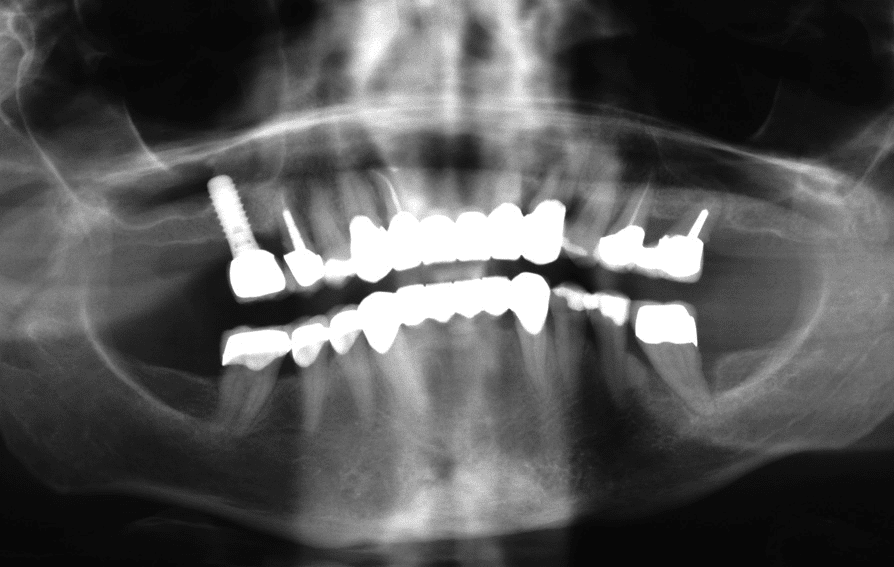
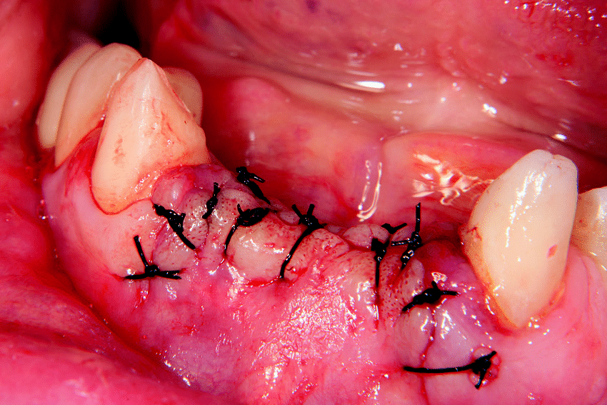
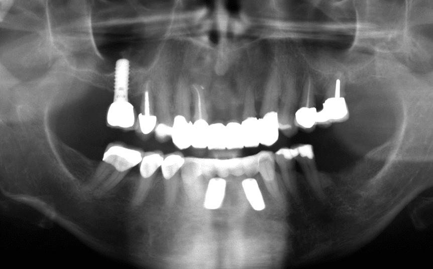
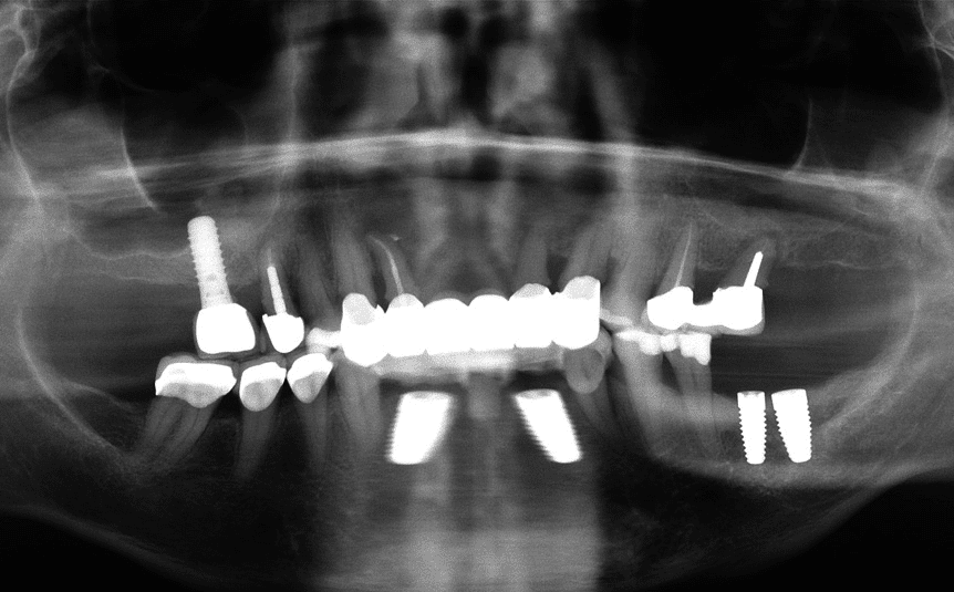
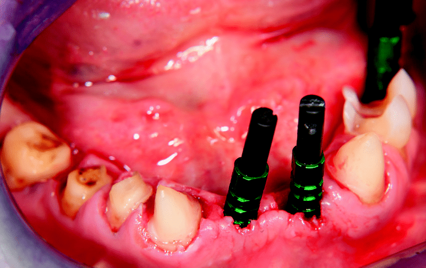
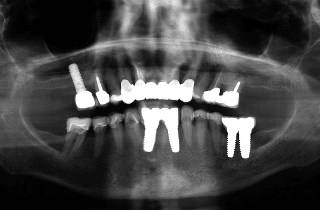
Fig.8: Wound closure after completion of the surgery. Fig.9: Clinical situation after two weeks and suture removal. Fig.10: OPG four weeks after bone splitting and placement of the two Zeramex XT implants in regions #32 and 42 (a) and three months after implant insertion in the posterior region (regions #36 and 37; b) 1a 3 6 9 1b 4 7 10a 2 5 8 10b 01 | case report sequent bone healing, two ceramic implants were to be placed in these regions. In addition, all remaining natural teeth required new prosthetic restoration with crowns.
Materials
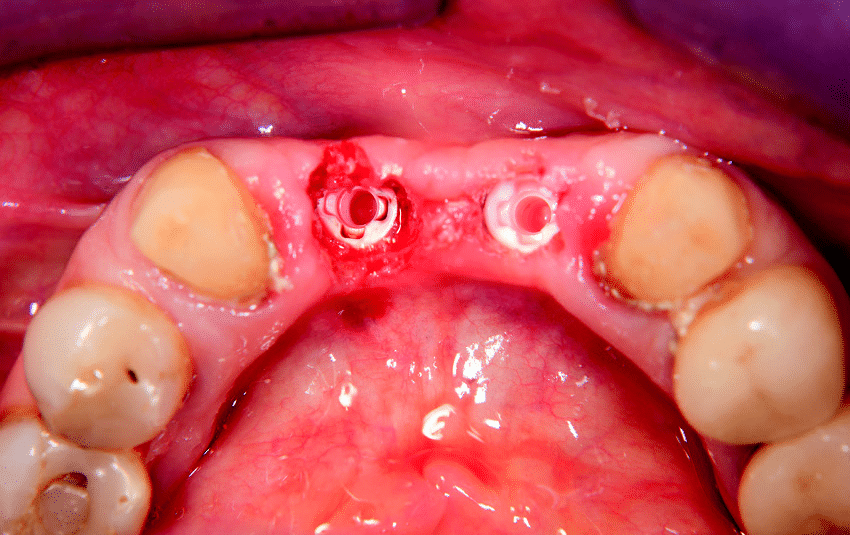
The implants used in this case were Zeramex XT implants (Dentalpoint) with diameters of 4.2mm (regular base) and 5.5mm (wide base) and a length of 10.0mm. In addition, maxresorb inject (botiss biomaterials) and a Jason membrane (botiss biomaterials) were used as part of the bone grafting procedure to cover the bony structures (Figs. 6–14).
Practical relevance
Alveolar ridge-splitting and expansion techniques are suitable and well-established methods for managing narrow residual bone with relatively minor effort.1 Different techniques and utilisation of various types of instruments are described in the literature, and only minor differences between them are reported regarding survival of the implants.2,3 Furthermore, study reviews have revealed a low rate of intra- and postoperative complications, such as buccal wall fracture, which requires careful preparation of the ridge and prudent selection of patients.4 Based on the clinical experience of the author, the technique employed for this case works particularly well in combination with ceramic implants owing to their high biocompatibility and material properties.
Conclusion
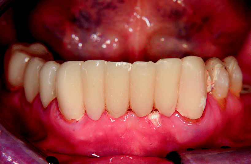
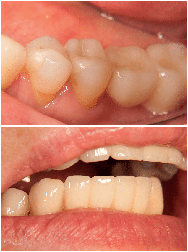
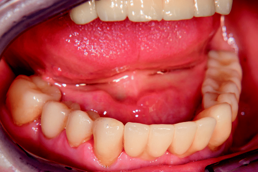
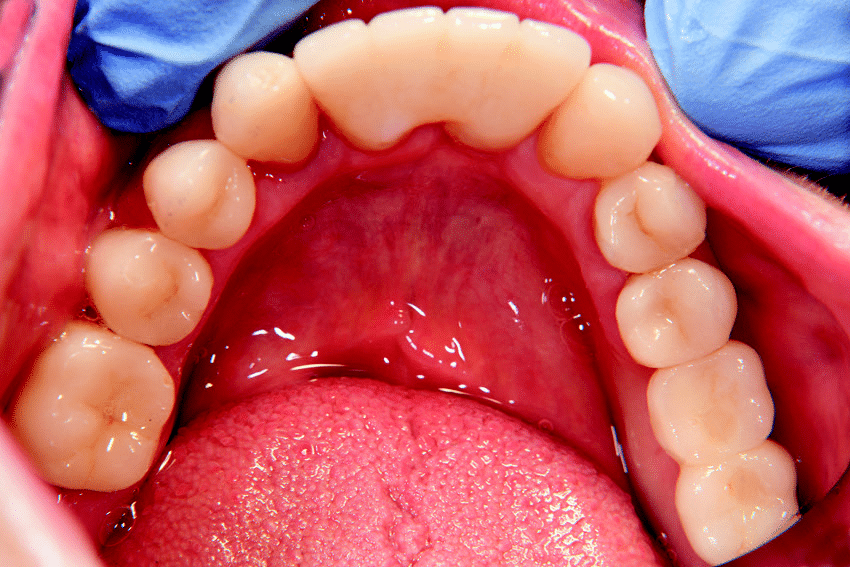
After more than ten years of working with two-piece ceramic implants and with more than 1,100 Zeramex implants of several types and generations having been placed, the author feels confident in using this technology in conjunction with ceramic implants restored with bridges. In his daily routine, the Zeramex XT implant with its internal geometry in combination with the carbon fibre-reinforced high-performance PEEK screw (Vicarbo screw) allows patient-specific prosthetic restorations, including bridgework and removable dentures, with reliable long-term success, including stable aesthetic outcomes (Figs. 15–19).
About the Doctor

Dr. Michael Leistner
Dr. Michael Leistner is a Germany based dentist specialized in dental implantology, focusing particularly on the use of ceramic implants. He runs a private practice in the city of Merzhausen in Germany.
Dr. Michael Leistner: info@dent-design.de

Contact
Want to know more about Zeramex?
Email us at mfoley@zeramexusa.com with your contact details and we will get in touch with you!
ZERAMEX BROCHURE
Click here to download the Zeramex sales brochure